X-ray Cultural Heritage Inspection
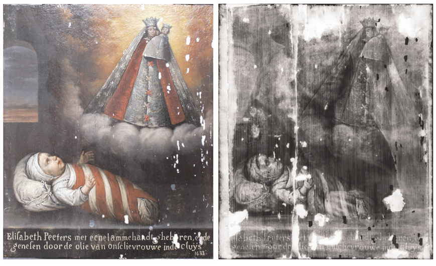
Through X-ray imaging, we can scrutinize the internal composition of an artwork, revealing an underlying sketch obscured beneath the visible subject. Beyond discerning the painting's history and authentication, artistic methodologies and variations in brightness and grayscale in the images indicate the material and density of pigments, contributing to a more nuanced understanding of the painting's historical context and providing essential criteria for authentication.1
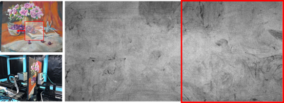
Using X-ray, the artwork is projected onto a detector. Automated equipment is employed to position and reconstruct the multiple images, enabling a comprehensive scan of the entire painting. The figure above illustrates the scanning of an oil painting sample on a five-axis X-ray platform, with the red mark indicating the position of the specific image.
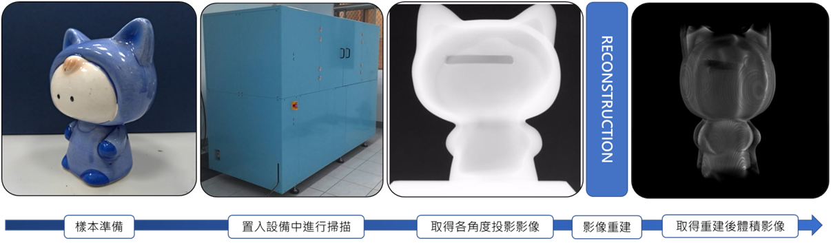
The sample in this case is a ceramic piggy bank with an internal hollow structure. The technique employed is CT imaging, capturing images at various angles within a semi-circular rotation to simplify the reconstruction process and reduce processing times. Using multiple methods, we reconstruct the image with dimensions of 640x480x512, in 8-bit grayscale.
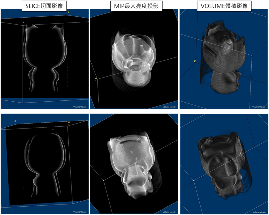
SLICE Image
The slice imaging technique reveals the internal structure and composition of an object, enabling observation from various angles irrespective of the original projection angle. Moving through different slices facilitates the identification of defects or points of interest, allowing for precise measurements of dimensions, positions, orientations, and material properties. This method enables comparisons of object surfaces or cross-sections.
Maximum Intensity Projection
The MIP projects three-dimensional spatial data onto a visualization plane. It adjusts the brightness of each voxel to display the maximum brightness on the projection plane. MIP effectively represents X-ray absorption values, highlighting even smaller readings, and provides a clear presentation of contrast and density variations.
VOLUME
Consider the voxel as density and assign them with grayscale values and coloring. This approach effectively visualizes the external appearance of the measured object or structural density variations across different sections. Volumetric images can undergo further mesh processing, serving as data foundations for reverse engineering or stress analysis.
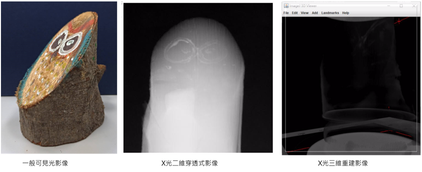
The above images depict wooden carved artworks. The eyes of this artwork are composed of glazed pigments, exhibiting higher density and X-ray absorption values. This characteristic is clearly discernible in X-ray imaging and computed tomography reconstructions.
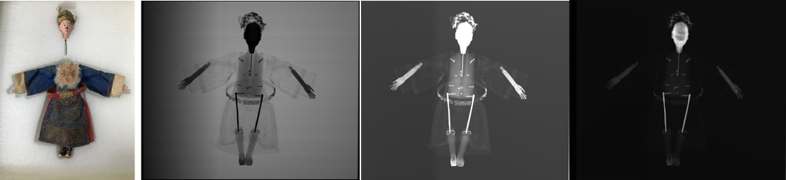
Exquisite puppet image:
The image on the right is the unprocessed raw file, while post-processing enhances the fabric and limb components, allowing for the observation of textile textures and internal structures. Moreover, the post-processing intensifies facial coloration, aiding in the identification of mineral or metallic pigments, indicating higher absorption rates.
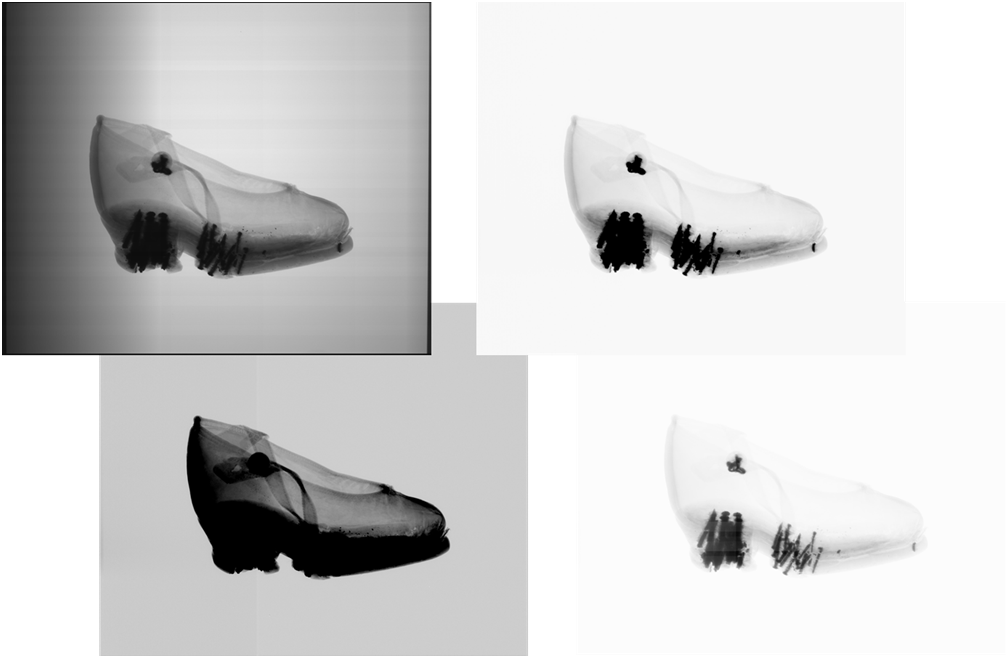
The image showcases footwear worn by women with ‘foot binding’ during the Qing Dynasty. Under X-ray examination, it reveals the structural reinforcement with metal components.
The top-left image represents the original raw file, while the bottom-left image depicts the processed version, we can identify the discernible patterns on the shoe surface. The top-right image shows the structural features of the footwear. The bottom-right image, focuses on enhancing the visibility of the metal nail components.
1 Schalm, Olivier & Vanbiervliet, Lies & Willems, Peter & Schepper, Peter. (2014). Radiography of paintings: Limitations of transmission radiography and exploration of emission radiography using phosphor imaging plates. Studies in Conservation. 59. 10-23. 10.1179/2047058413Y.0000000088.